
How Does a Transthoracic Echocardiogram Work?
A transthoracic echocardiogram bounces high-frequency sound off your heart from outside the chest and turns the echoes into moving pictures, while Doppler reads the speed and direction of blood through the valves. No radiation. Nothing goes inside you, and there's usually no prep. That makes it the natural first look when a symptom, a murmur, or suspected heart failure raises a specific question. A clearly abnormal study is often enough to guide the next move. A clean one is sometimes only the first step.


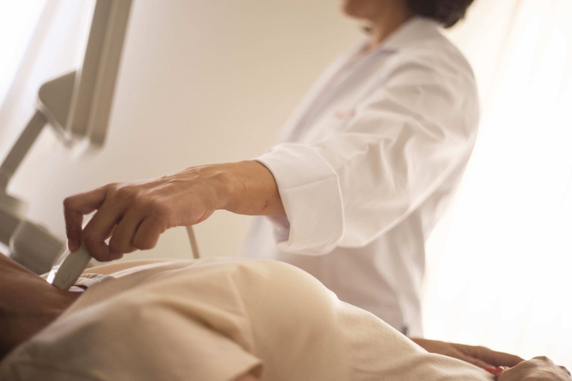
In one sitting, a transthoracic echocardiogram (TTE) shows how big your heart's chambers are, how well the muscle squeezes, whether the valves open and close properly, and how blood is moving. That's why it's the default first imaging test when something points to structural trouble. What it can't give you is a blanket all-clear. It misses some problems often enough that a normal result means the most when your symptoms and risk agree with it.
Gel, a probe, and the gaps between your ribs
The test itself is simple. A sonographer spreads gel on your chest and moves a handheld transducer over several spots, often with you lying on your left side. The probe sends out sound and listens for what bounces back from the boundaries between blood, muscle, and valve tissue, and a computer turns those echoes into still and moving images. A standard exam usually takes 30 to 60 minutes.
The hard part is getting sound to the heart at all. Ribs block it and air-filled lung scatters it, so the probe works through a few natural openings: beside the breastbone, at the heart's tip, below the ribcage, and in the notch above the breastbone. Each view shows different structures. The view from the tip lines the beam up with blood flowing through the valves, which matters because Doppler underestimates speed when the angle is off. The view from under the ribs uses the liver as a path when the lungs get in the way.
So image quality varies a lot between people. Obesity, lung disease, a ventilator, or chest tubes can all degrade the pictures. In one study of severe chest trauma, most transthoracic images were suboptimal.
Safety is the easy part. Ultrasound doesn't use radiation, and MedlinePlus lists no known risks for the routine external exam. The FDA notes that ultrasound energy can slightly warm tissue, which is why operators use the lowest exposure that still gives a diagnostic picture.
Shape and flow in the same exam
An echo is more than a photograph. The images show anatomy, and Doppler adds how the blood behaves. Red blood cells moving toward or away from the probe shift the frequency of the returning sound, and the machine turns that shift into speed and direction. Speed across a narrowed valve can then be converted into a pressure difference. That's how an echo grades a stiff valve without a catheter. The American Society of Echocardiography defines a complete adult exam as combining all of these, with extras like contrast, strain, or 3D added when useful.
| Part of the exam | What it looks at | Why it matters |
|---|---|---|
| 2D and M-mode imaging | Chamber size, wall thickness, valve leaflets, how each wall segment moves, ejection fraction | Shows a weak or enlarged heart, damage from a past heart attack, or fluid around the heart |
| Color Doppler | Direction and pattern of blood flow, painted over the image | Reveals leaky valves and abnormal jets and helps grade how bad a leak is |
| Spectral Doppler | Exact blood speeds at one spot or along a line through a valve | Estimates pressure gradients across narrowed valves and shows how the heart fills |
| Contrast-enhanced TTE | IV microbubble agent or agitated saline | Sharpens the heart's inner border when images are poor; saline can show abnormal right-to-left passage of bubbles |
Some of these measurements also tell you how seriously to take a finding. In large cohorts, an enlarged ventricle and wall segments that don't move normally were each linked to more heart failure hospitalizations and deaths. That's an association, not proof the echo itself changes the heart's course.
Contrast is the one part with a formal warning. Some FDA labels carry a boxed warning for uncommon serious heart and lung reactions, so these agents are given where resuscitation equipment is on hand. In a nationwide claims analysis, people who got contrast had lower odds of dying within two days than matched people who didn't. That's observational, but it fits with serious harm being rare.
Best used when the answer will change something
All that information is only worth having if someone will act on it, and guidelines put TTE first in line for a defined set of problems. The 2022 heart failure guideline from the American Heart Association, American College of Cardiology, and Heart Failure Society of America gives it the strongest recommendation for the first evaluation of suspected or new heart failure. After an acute coronary syndrome, it's standard for checking pumping function and looking for complications or a clot in the ventricle. It's also the starting test for a new murmur, known valve disease, unexplained shock, and a stroke that may have come from the heart.
When there's a real question, the answer often matters. At one academic medical center, TTE led to an active change in care in about a third of cases and confirmed the existing plan in nearly half. In stroke patients, finding a high-risk cardiac source on TTE was linked to fewer recurrent stroke hospitalizations over the next year, plausibly because it led to targeted treatment. That's an observational link, not a trial result.
If you already have a valve problem, TTE is how it's tracked. The American Heart Association's ordering guide scales the interval to severity, from every 3 to 5 years for mild disease to every 6 to 12 months for severe aortic stenosis without symptoms. New symptoms reset that clock.
Echoes ordered with no question attached don't earn much. The same heart failure guideline says a repeat scan just to recheck function isn't useful when nothing has changed. And hospitals that order echoes most liberally for heart attack patients haven't shown better survival. Just longer stays and higher costs.
Trust a positive echo more than a negative one
For several specific questions, including endocarditis, direct signs of acute tears in the main artery near the heart, and contrast TTE for right-to-left shunts, meta-analyses show positive findings tend to be more reliable than negative ones. When it shows a growth on a valve or a flap in the aorta, the finding is usually real.
Misses are the weak side, and some are large. For infected valve growths, modern TTE caught only about 61% of those seen on a transesophageal echocardiogram (TEE), where the probe goes down the esophagus right behind the heart. In patients with ischemic heart disease, TTE found about a quarter of the ventricular clots that contrast cardiac MRI found. Clots in the left atrial appendage and some lesions at the heart's tip are known blind spots.
So a clean echo means different things in different situations. If you don't have an artificial valve, a clearly negative TTE does substantially lower the odds of endocarditis. But with bacteria in the blood and an artificial valve, or a symptom that still hasn't been explained, a normal echo is a reason for the next test, whether TEE, CT, or cardiac MRI. Not a reason to stop.
Sudden tearing chest or back pain is a separate matter. That needs emergency evaluation, because TTE alone misses too many aortic dissections to rule one out.
If it's safe, why not get one anyway?
It's the obvious question for a proactive person. The Tromsø trial in Norway tested it, randomizing 6,861 middle-aged adults to a screening echo or usual care and following them for 15 years.
That's not because screening finds nothing. In the OxVALVE study of adults 65 and older, it turned up previously unknown valve lesions in about half, though most were mild, and a stethoscope isn't a reliable backstop: in one primary-care study it caught fewer than half of significant valve lesions. The gap is what happens next. For many early, mild findings there's no proven treatment that changes their course, so knowing sooner hasn't translated into living longer.
That points the test at specific people rather than everyone. Breathlessness on stairs, new fatigue, a murmur someone heard, abnormal heart biomarkers, or symptoms at an older age that you've been writing off are where an echo tends to change what happens. And if you turn out to have moderate or severe valve disease, finding it starts the surveillance that times treatment, even though screening everyone hasn't shown a survival gain. Researchers have developed AI-read ECG tools to flag who is more likely to have undiagnosed structural disease.
A TTE is a safe first look at how your heart is built and how it moves blood. It's at its best answering a specific question, and sometimes its answer opens the workup rather than closing it. That would change if a trial showed that echo screening of low-risk adults, or of a well-chosen higher-risk group, prevents deaths or hospitalizations, or if better imaging methods cut missed findings in a way that improves outcomes, not just pictures.